Pathology is one of the most important subjects for USMLE Step 1 because it connects basic science to clinical disease. If you understand pathology well, physiology, pharmacology, immunology, microbiology, and biochemistry become easier to apply inside clinical vignettes.
Many students try to memorize pathology as a long list of diseases. That is the wrong approach. Step 1 does not simply ask, “What is the diagnosis?” It asks whether you understand the mechanism behind the disease.
The most important pathology concepts for Step 1 include cell injury, inflammation, tissue repair, hemodynamic disorders, neoplasia, immunopathology, genetic disease, environmental pathology, and organ-system pathology.
This guide breaks down the pathology topics you should master first, how Step 1 tests them, and how to study them using clinical reasoning instead of random memorization.
Free Worksheet
Download the General Pathology Masterclass Worksheet
Use this worksheet to organize high-yield general pathology concepts, connect mechanisms to clinical vignettes, and strengthen your USMLE Step 1 pathology foundation.
Get the Free WorksheetFree USMLE Training
Free USMLE Step 1 Bootcamp
Join our free USMLE masterclass where we break down high-yield concepts, NBME strategies, and clinical reasoning systems used by thousands of medical students and IMGs.
Reserve My SpotWhy Pathology Matters So Much for USMLE Step 1
Pathology is the language of disease. Step 1 uses pathology to test whether you can connect normal physiology to abnormal physiology, then apply that mechanism to a patient vignette.
If your pathology foundation is weak, you may recognize isolated facts but struggle when a question combines symptoms, labs, histology, and pathophysiology.
The Big Rule
Do not study pathology by memorizing disease names only. Study the mechanism, the clinical pattern, the lab abnormality, and the histology clue together.
| Pathology Area | Why It Matters | How Step 1 Tests It |
|---|---|---|
| Cell Injury | Foundation for ischemia, toxins, necrosis, and organ damage | Mechanisms of reversible injury, irreversible injury, free radicals, and ATP depletion |
| Inflammation | Explains infection, autoimmune disease, granulomas, and healing | Cytokines, leukocyte adhesion, neutrophils, macrophages, and chronic inflammation |
| Neoplasia | Core cancer biology tested across organ systems | Tumor suppressors, oncogenes, paraneoplastic syndromes, invasion, and metastasis |
| Hemodynamics | Explains thrombosis, embolism, edema, infarction, and shock | Virchow triad, pulmonary embolism, DIC, infarct patterns, and shock types |
Cell Injury, Necrosis, and Apoptosis
Cell injury is one of the highest-yield pathology foundations for Step 1. You need to understand what happens when cells lose oxygen, lose ATP, accumulate calcium, generate free radicals, or experience membrane damage.
High-Yield Cell Injury Concepts
- Reversible injury: cellular swelling, fatty change, membrane blebs, ribosomal detachment.
- Irreversible injury: severe membrane damage, mitochondrial dysfunction, nuclear changes.
- ATP depletion: failure of Na/K ATPase, cellular swelling, anaerobic glycolysis, lactic acidosis.
- Free radical injury: lipid peroxidation, protein damage, DNA damage.
- Calcium influx: activates phospholipases, proteases, endonucleases, and ATPases.
Necrosis Patterns You Must Know
| Necrosis Type | Classic Association | Step 1 Clue |
|---|---|---|
| Coagulative necrosis | Ischemic infarction in most solid organs | Architecture preserved, dead tissue remains firm |
| Liquefactive necrosis | Brain infarcts and abscesses | Enzymatic digestion creates liquid material |
| Caseous necrosis | Tuberculosis and fungal infections | Cheese-like necrosis inside granulomas |
| Fat necrosis | Acute pancreatitis or breast trauma | Calcium soap formation and chalky white deposits |
| Fibrinoid necrosis | Immune-mediated vascular injury | Bright pink vessel wall deposition |
Step 1 Trap
Apoptosis is programmed cell death without inflammation. Necrosis causes membrane rupture and inflammation. That difference is tested repeatedly.
Acute and Chronic Inflammation
Inflammation is tested across microbiology, immunology, pathology, rheumatology, and organ-system disease. You must know the cells, cytokines, vascular changes, and timing.
Acute Inflammation
Acute inflammation is usually dominated by neutrophils. It involves vasodilation, increased vascular permeability, leukocyte adhesion, migration, chemotaxis, and phagocytosis.
- Neutrophils: first responders in acute bacterial infection.
- Histamine: vasodilation and increased vascular permeability.
- TNF-alpha and IL-1: endothelial activation and fever.
- IL-8: neutrophil chemotaxis.
- C5a: chemotaxis and neutrophil activation.
Chronic Inflammation
Chronic inflammation is dominated by macrophages, lymphocytes, plasma cells, tissue destruction, and fibrosis. Granulomatous inflammation is especially high yield.
Granuloma Rule
Granulomas form when macrophages transform into epithelioid histiocytes to wall off material that is difficult to eradicate, such as tuberculosis, fungi, foreign bodies, or sarcoidosis.
Wound Healing and Tissue Repair
Wound healing questions often test timing. Step 1 may give a wound biopsy at a specific day and ask which cell type or process is most active.
| Timeline | Main Process | Key Cell or Feature |
|---|---|---|
| First 24 hours | Hemostasis and early inflammation | Platelets and neutrophils |
| 1 to 3 days | Acute inflammation | Neutrophils dominate |
| 3 to 7 days | Granulation tissue formation | Macrophages, fibroblasts, angiogenesis |
| 1 to 2 weeks | Collagen deposition | Fibroblasts and type III collagen |
| Weeks to months | Remodeling | Type III collagen replaced by type I collagen |
Also know impaired wound healing causes, including diabetes mellitus, vitamin C deficiency, zinc deficiency, corticosteroids, infection, poor perfusion, and malnutrition.
Hemodynamics, Thrombosis, Embolism, and Shock
Hemodynamics is one of the most testable pathology areas because it links pathology to cardiology, pulmonology, renal disease, obstetrics, surgery, and emergency medicine.
Virchow Triad
- Endothelial injury: atherosclerosis, vasculitis, hypertension, trauma.
- Abnormal blood flow: stasis, turbulence, atrial fibrillation, aneurysm.
- Hypercoagulability: Factor V Leiden, malignancy, pregnancy, OCPs, nephrotic syndrome.
Shock Types
| Shock Type | Classic Cause | Key Concept |
|---|---|---|
| Hypovolemic shock | Hemorrhage, dehydration, burns | Low preload and low cardiac output |
| Cardiogenic shock | Myocardial infarction, severe heart failure | Pump failure |
| Septic shock | Severe infection | Distributive shock with inflammatory vasodilation |
| Anaphylactic shock | IgE-mediated mast cell degranulation | Vasodilation, bronchospasm, urticaria |
| Neurogenic shock | Spinal cord injury | Loss of sympathetic tone |
Neoplasia and Cancer Biology
Cancer biology is heavily tested on Step 1. You must understand oncogenes, tumor suppressor genes, metastasis, grading, staging, paraneoplastic syndromes, and cancer-associated mutations.
High-Yield Cancer Genes
- TP53: DNA damage checkpoint, apoptosis, Li-Fraumeni syndrome.
- RB: G1 to S checkpoint, retinoblastoma, osteosarcoma.
- APC: Wnt pathway regulation, familial adenomatous polyposis.
- BRCA1 and BRCA2: DNA repair, breast and ovarian cancer.
- RAS: signal transduction, many solid tumors.
- MYC: Burkitt lymphoma translocation.
- BCR-ABL: chronic myeloid leukemia.
Step 1 Cancer Rule
Grading describes how abnormal the tumor looks under the microscope. Staging describes how far the tumor has spread. Staging is usually more important for prognosis.
High-Yield Step 1 Resource
SmashUSMLE High Yield Step 1 Book
If pathology feels overwhelming, use the SmashUSMLE High Yield Step 1 Book to organize disease mechanisms, clinical patterns, and board-style reasoning.
- Strengthen high-yield Step 1 foundations
- Review pathology mechanisms more efficiently
- Connect disease patterns to NBME-style questions
- Use alongside SmashUSMLE, NBME review, and QBank practice

Immunopathology and Hypersensitivity
Immunopathology is frequently tested because it explains autoimmune disease, vasculitis, glomerulonephritis, transplant rejection, allergic reactions, and immune deficiencies.
| Type | Mechanism | Classic Examples |
|---|---|---|
| Type I | IgE-mediated mast cell degranulation | Anaphylaxis, asthma, allergic rhinitis |
| Type II | Antibody-mediated cell or receptor damage | Goodpasture syndrome, autoimmune hemolytic anemia, Graves disease, myasthenia gravis |
| Type III | Immune complex deposition | SLE, serum sickness, poststreptococcal glomerulonephritis |
| Type IV | T-cell mediated delayed hypersensitivity | Contact dermatitis, TB skin test, type 1 diabetes, granulomas |
Genetic and Pediatric Pathology
Genetic pathology questions often test inheritance patterns, trinucleotide repeats, imprinting, chromosomal abnormalities, tumor syndromes, and pediatric disease mechanisms.
High-Yield Genetic Pathology Topics
- Autosomal dominant vs autosomal recessive inheritance
- X-linked recessive disease patterns
- Mitochondrial inheritance
- Trinucleotide repeat expansion and anticipation
- Genomic imprinting
- Down syndrome, Turner syndrome, Klinefelter syndrome
- Marfan syndrome, Ehlers-Danlos syndrome, osteogenesis imperfecta
- Lysosomal storage diseases
Environmental and Nutritional Pathology
Environmental pathology is high yield because Step 1 loves toxins, occupational exposures, smoking-related disease, alcohol-related disease, and nutritional deficiencies.
- Lead: abdominal pain, neuropathy, anemia, basophilic stippling.
- Carbon monoxide: tissue hypoxia with normal PaO2, cherry-red appearance.
- Asbestos: pleural plaques, mesothelioma, bronchogenic carcinoma.
- Silica: upper-lobe lung disease, increased TB risk.
- Alcohol: fatty liver, hepatitis, cirrhosis, pancreatitis.
- Vitamin C deficiency: poor wound healing and bleeding gums.
- Vitamin B12 deficiency: megaloblastic anemia and neurologic deficits.
Histology and Pathology Image Recognition
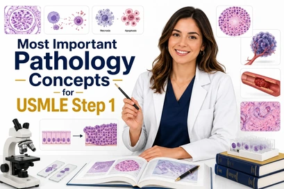
Step 1 pathology images are not random. They usually test classic visual patterns that connect directly to a disease mechanism.
Images You Should Recognize
- Reed-Sternberg cells in Hodgkin lymphoma
- Starry-sky pattern in Burkitt lymphoma
- Caseating granulomas in tuberculosis
- Noncaseating granulomas in sarcoidosis
- Rouleaux formation in multiple myeloma
- Smudge cells in CLL
- Auer rods in acute myeloid leukemia
- Psammoma bodies in papillary thyroid carcinoma
- Koilocytes in HPV infection
- Fat necrosis with saponification in pancreatitis
Image Strategy
Do not just memorize the picture. Ask what mechanism created the finding. That is how Step 1 turns an image into a reasoning question.
How to Study Pathology for USMLE Step 1
The best way to study pathology is to move from mechanism to clinical pattern to question application.
The SmashUSMLE Pathology Study Method
- Start with the mechanism: What is the disease process?
- Add the clinical picture: What symptoms and signs appear?
- Add the labs: What lab pattern confirms the mechanism?
- Add the image: What histology or gross pathology finding fits?
- Apply it to questions: Use QBank and NBME-style practice.
If you miss pathology questions, do not just read the explanation and move on. Write down why you missed it. Was it a content gap, mechanism gap, image recognition issue, or distractor trap?
Student Success Story
⭐ 4.8 Google Rating | 120+ ReviewsSee How Dr. Evelyn Passed Step 1 and Step 2
Dr. Evelyn’s story shows how structured clinical reasoning, NBME-focused review, and disciplined preparation can help students move forward with confidence.
Want to learn the same clinical reasoning system used by SmashUSMLE students?
Join Free BootcampNeed Help Mastering Pathology for Step 1?
If pathology feels overwhelming, the problem is usually not effort. The problem is structure. You need to know which concepts matter most, how Step 1 tests them, and how to apply them inside clinical vignettes.
SmashUSMLE Reviews helps students use high-yield review, clinical reasoning, QBank practice, NBME weak-area analysis, and one-on-one tutoring to prepare smarter.
FAQ: Most Important Pathology Concepts for USMLE Step 1
What are the most important pathology concepts for USMLE Step 1?
The most important pathology concepts include cell injury, necrosis, apoptosis, inflammation, wound healing, hemodynamics, thrombosis, embolism, shock, neoplasia, immunopathology, genetic disease, and histology recognition.
Is pathology high yield for Step 1?
Yes. Pathology is one of the highest-yield subjects for Step 1 because it connects basic science mechanisms to clinical disease presentations.
How should I study pathology for Step 1?
Study pathology by learning the mechanism first, then connecting it to symptoms, labs, histology, and clinical vignette patterns. Do not rely only on memorization.
Do I need to know histology for Step 1 pathology?
Yes. You should recognize classic histology findings such as granulomas, Reed-Sternberg cells, Auer rods, smudge cells, starry-sky pattern, psammoma bodies, and koilocytes.
What is the best way to review missed pathology questions?
For each missed question, identify whether the problem was a content gap, mechanism gap, image recognition issue, or distractor trap. Then review that specific weakness before doing more questions.
Can SmashUSMLE help with pathology for Step 1?
Yes. SmashUSMLE helps students review high-yield pathology, connect mechanisms to clinical reasoning, use QBank practice effectively, and target NBME weak areas.
Ready to Improve Your USMLE Scores?
Step 1 preparation becomes easier when you stop memorizing randomly and start using a system. Join thousands of medical students and IMGs using SmashUSMLE’s clinical reasoning system to prepare for Step 1, Step 2 CK, and Step 3.