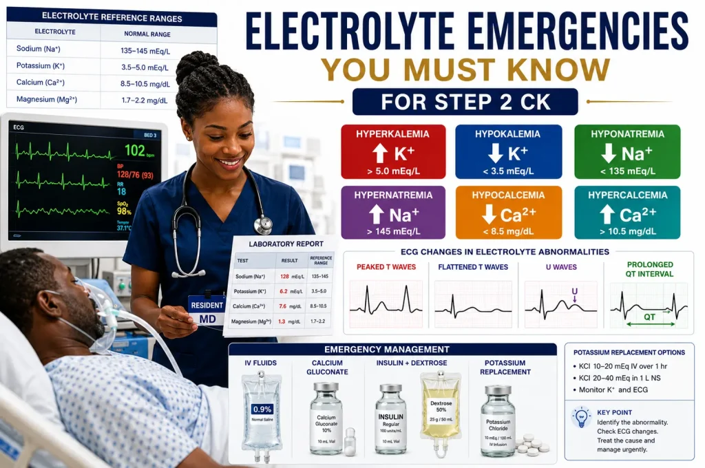

Electrolyte emergencies on Step 2 CK are not just about memorizing normal lab values. The exam wants to know if you can recognize danger, stabilize the patient, and choose the next best step before the patient crashes.
That means you must quickly connect the lab abnormality to the clinical picture. Hyperkalemia with ECG changes is treated differently from mild asymptomatic hyperkalemia. Severe symptomatic hyponatremia is treated differently from chronic mild hyponatremia.
Step 2 CK often hides the diagnosis inside a vignette. The patient may have renal failure, vomiting, diarrhea, diuretic use, DKA, malignancy, seizures, confusion, weakness, tetany, or arrhythmias.
In this guide, we will break down the electrolyte emergencies you must know for Step 2 CK using pattern recognition, emergency treatment, and USMLE-style questions.
Table of Contents
Free USMLE Step 2 CK Bootcamp
Join our free USMLE masterclass where we break down high-yield concepts, NBME strategies, and clinical reasoning systems used by thousands of medical students and IMGs.
Electrolyte Emergencies on Step 2 CK: The High-Yield Framework
When Step 2 CK gives you an abnormal electrolyte, do not panic. Ask three questions first.
- Is the patient symptomatic? Look for seizures, confusion, coma, weakness, tetany, arrhythmia, or shock.
- Are there ECG changes? ECG changes make potassium and calcium abnormalities much more urgent.
- Is the abnormality acute or chronic? Acute sodium shifts are more dangerous and treated more aggressively.
The Big Four Electrolytes
The highest-yield electrolytes for Step 2 CK are potassium, sodium, calcium, and magnesium. Each one has a classic emergency presentation.
- Potassium: arrhythmias, weakness, peaked T waves, U waves.
- Sodium: seizures, confusion, coma, cerebral edema, osmotic demyelination risk.
- Calcium: tetany, seizures, stones, bones, groans, shortened or prolonged QT.
- Magnesium: torsades de pointes, seizures in preeclampsia, hyporeflexia from toxicity.
Potassium Emergencies on Step 2 CK
Potassium emergencies are heavily tested because they cause life-threatening arrhythmias. The key is to separate hyperkalemia from hypokalemia and know the immediate next step.
Hyperkalemia
Hyperkalemia commonly occurs in renal failure, ACE inhibitor use, ARB use, spironolactone use, potassium supplements, tissue breakdown, tumor lysis syndrome, rhabdomyolysis, and metabolic acidosis.
The ECG clues are the key. Look for peaked T waves, PR prolongation, QRS widening, sine-wave pattern, ventricular arrhythmias, or cardiac arrest.
Hyperkalemia Treatment Sequence
- IV calcium gluconate if ECG changes or severe symptoms are present.
- Insulin plus dextrose to shift potassium into cells.
- Albuterol can also shift potassium intracellularly.
- Remove potassium with loop diuretics, potassium binders, or dialysis.
Hypokalemia
Hypokalemia usually presents with muscle weakness, cramps, ileus, arrhythmias, and ECG changes. Common causes include vomiting, diarrhea, loop diuretics, thiazides, hyperaldosteronism, insulin therapy, and beta-agonist use.
ECG findings include flattened T waves, ST depression, prominent U waves, and prolonged QU interval. Severe hypokalemia can cause ventricular arrhythmias.
A 67-year-old man with chronic kidney disease presents with weakness and palpitations. Potassium is 6.9 mEq/L. ECG shows peaked T waves and widening of the QRS complex.
What is the most appropriate next step?
A. Albuterol nebulization only
B. IV calcium gluconate
C. Oral potassium binder only
D. Repeat potassium level in 6 hours
Correct Answer: B. IV calcium gluconate
This patient has severe hyperkalemia with ECG changes. The first step is IV calcium gluconate to stabilize the myocardium. After that, insulin with dextrose and albuterol can shift potassium intracellularly. Potassium removal is still required afterward.
High Yield Clinical Pearl: ECG changes in hyperkalemia mean calcium first.
Download the Free Nephrology Masterclass Worksheet
Get the high-yield nephrology worksheet used by SmashUSMLE students to review fluids, electrolytes, acid-base disorders, renal failure, diuretics, and emergency management patterns.
Download Free WorksheetSodium Emergencies on Step 2 CK
Sodium questions test whether you understand brain swelling and safe correction. The danger in acute hyponatremia is cerebral edema. The danger in overcorrecting chronic hyponatremia is osmotic demyelination syndrome.
Hyponatremia
Hyponatremia can present with headache, nausea, confusion, seizures, coma, and respiratory arrest. Severe neurologic symptoms matter more than the exact sodium number.
Causes include SIADH, diuretics, heart failure, cirrhosis, nephrotic syndrome, adrenal insufficiency, primary polydipsia, vomiting, diarrhea, and hypotonic IV fluids.
Symptomatic Hyponatremia Treatment
If the patient has seizures, coma, or severe neurologic symptoms, treat with hypertonic saline. Do not choose fluid restriction for a patient actively seizing from severe hyponatremia.
Hypernatremia
Hypernatremia usually reflects water loss greater than sodium loss. Patients may have dehydration, altered mental status, weakness, irritability, seizures, or coma.
Common causes include diabetes insipidus, poor access to water, osmotic diuresis, fever, diarrhea, burns, and excessive sodium administration.
If the patient is hypovolemic and unstable, restore intravascular volume first with isotonic saline. Then correct free water deficit carefully.
A 45-year-old woman is brought to the emergency department after a generalized tonic-clonic seizure. She has been taking hydrochlorothiazide. Serum sodium is 116 mEq/L.
What is the best next step?
A. Fluid restriction only
B. Hypertonic saline
C. Oral sodium tablets only
D. Tolvaptan as first-line therapy
Correct Answer: B. Hypertonic saline
This patient has severe symptomatic hyponatremia with seizure. Hypertonic saline is the next step. Fluid restriction may be used in stable SIADH, but not in acute severe neurologic symptoms.
High Yield Clinical Pearl: Severe hyponatremia plus seizure means hypertonic saline.
Calcium Emergencies on Step 2 CK
Calcium abnormalities show up with neuromuscular symptoms, psychiatric changes, kidney stones, abdominal pain, bone pain, and ECG changes.
Hypocalcemia
Hypocalcemia causes paresthesias, muscle cramps, tetany, seizures, laryngospasm, Chvostek sign, Trousseau sign, and prolonged QT interval.
Common causes include hypoparathyroidism after thyroid surgery, vitamin D deficiency, chronic kidney disease, pancreatitis, sepsis, massive transfusion, and hypomagnesemia.
Severe symptomatic hypocalcemia requires IV calcium gluconate.
Hypercalcemia
Hypercalcemia can cause constipation, abdominal pain, kidney stones, polyuria, dehydration, confusion, weakness, shortened QT interval, and coma.
The most common causes are primary hyperparathyroidism and malignancy. In a severely symptomatic patient, initial treatment is aggressive IV isotonic saline.
After fluids, calcitonin can provide short-term reduction, while bisphosphonates provide longer-term control, especially in malignancy-associated hypercalcemia.
A 59-year-old man with metastatic lung cancer presents with confusion, constipation, weakness, and polyuria. Calcium is 14.8 mg/dL. Mucous membranes are dry.
What is the best initial treatment?
A. Aggressive IV normal saline
B. Fluid restriction
C. Immediate thyroidectomy
D. Oral calcium supplementation
Correct Answer: A. Aggressive IV normal saline
This patient has severe symptomatic hypercalcemia, likely from malignancy. The initial treatment is IV isotonic saline to correct dehydration and increase renal calcium excretion. Calcitonin and bisphosphonates can be added after volume resuscitation.
High Yield Clinical Pearl: Severe hypercalcemia is treated first with IV fluids.
Magnesium Emergencies on Step 2 CK
Magnesium is easy to overlook, but Step 2 CK likes it because it connects to torsades de pointes, refractory hypokalemia, and preeclampsia treatment.
Hypomagnesemia
Hypomagnesemia can cause tremor, weakness, seizures, arrhythmias, torsades de pointes, hypokalemia, and hypocalcemia.
Common causes include alcoholism, malnutrition, diarrhea, proton pump inhibitor use, aminoglycosides, amphotericin B, cisplatin, and diuretics.
Hypermagnesemia
Hypermagnesemia is usually caused by renal failure or excessive magnesium administration. Symptoms include nausea, flushing, hypotension, bradycardia, hyporeflexia, respiratory depression, and cardiac arrest.
Magnesium toxicity is treated with IV calcium gluconate, supportive care, stopping magnesium, and dialysis if severe renal failure is present.
Torsades de Pointes
Torsades is a polymorphic ventricular tachycardia associated with prolonged QT. The treatment is IV magnesium sulfate, even if the serum magnesium level is normal.
A 31-year-old woman has recurrent episodes of syncope. ECG shows polymorphic ventricular tachycardia with twisting of the QRS complexes around the baseline. QT interval is prolonged.
What is the best treatment?
A. IV magnesium sulfate
B. Oral calcium carbonate
C. Potassium restriction
D. Verapamil
Correct Answer: A. IV magnesium sulfate
This patient has torsades de pointes. The treatment is IV magnesium sulfate. Also correct hypokalemia, stop QT-prolonging medications, and treat instability if present.
High Yield Clinical Pearl: Torsades equals magnesium.
Electrolyte Emergency Table for Step 2 CK
| Emergency | Classic Clues | ECG Finding | Next Best Step |
|---|---|---|---|
| Hyperkalemia | Renal failure, weakness, palpitations | Peaked T waves, widened QRS | IV calcium if ECG changes, then insulin/dextrose |
| Hypokalemia | Diuretics, vomiting, diarrhea, weakness | U waves, flattened T waves | Potassium replacement and magnesium correction |
| Symptomatic Hyponatremia | Seizures, confusion, coma | Usually nonspecific | Hypertonic saline |
| Hypernatremia | Dehydration, diabetes insipidus, altered mental status | Usually nonspecific | Volume resuscitation if unstable, then free water correction |
| Hypocalcemia | Tetany, seizures, Chvostek, Trousseau | Prolonged QT | IV calcium gluconate if severe or symptomatic |
| Hypercalcemia | Stones, bones, groans, psychiatric symptoms | Shortened QT | IV normal saline first |
| Hypomagnesemia | Alcohol use, diarrhea, refractory hypokalemia | Prolonged QT, torsades risk | Magnesium replacement |
| Hypermagnesemia | Renal failure, magnesium therapy, hyporeflexia | Bradycardia, conduction delay | IV calcium gluconate if toxic |
See How SmashUSMLE Helps Students Think Clinically
Electrolyte questions feel hard when students memorize isolated facts. They become easier when you connect the abnormal lab to the symptom, ECG, and emergency treatment.
If electrolyte questions feel confusing because every answer choice looks reasonable, clinical reasoning can help you move faster and avoid dangerous traps.
Join Free BootcampNeed Help Mastering Step 2 CK Emergency Questions?
If you're struggling to improve NBME performance or connect concepts clinically, the SmashUSMLE Masterclass was built to help students develop stronger clinical reasoning, high-yield retention, and test-day decision making.
FAQ: Electrolyte Emergencies on Step 2 CK
What electrolyte emergency is most important for Step 2 CK?
Hyperkalemia is one of the most important electrolyte emergencies because it can cause fatal arrhythmias. If ECG changes are present, give IV calcium first.
When do you give hypertonic saline on Step 2 CK?
Give hypertonic saline for severe symptomatic hyponatremia, especially when the patient has seizures, coma, or severe neurologic symptoms.
What is the first treatment for severe hypercalcemia?
The first treatment is aggressive IV isotonic saline. Calcitonin and bisphosphonates may be added after volume resuscitation.
What electrolyte treats torsades de pointes?
Torsades de pointes is treated with IV magnesium sulfate, even if the measured magnesium level is normal.
Why does hypomagnesemia cause refractory hypokalemia?
Low magnesium increases renal potassium wasting. Therefore, potassium may not correct until magnesium is replaced.
Ready to Improve Your USMLE Scores?
Join thousands of medical students and IMGs using SmashUSMLE’s clinical reasoning system to prepare for Step 1, Step 2 CK, and Step 3.